Cell: History and cell theory, Discovery, Features, Functional characteristics, Size and shape
Cell

Micrograph the scanning electron microscope cell Escherichia coli.
A cell (from the Latin cellula, a diminutive of cella, hollow) is the unit morphological and functional all living beings. In fact, the cell is the smallest element that can be considered alive. In this way, can be classified living organisms according to the number which have: if you only have one, are called unicellular (such as the protozoa or bacteria, microscopic organisms), if they have more, they are called multicellular. In the latter the number of cells varies from a few hundred, as in some nematodes, hundreds of billions (10 14), as in the case of the human being. The cells usually have a size of 10 μm and a mass of 1 ng, while cells are much greater.
The cell theory, proposed in 1839 by Matthias Jakob Schleiden and Theodor Schwann, postulates that all organisms are composed of cells and all cells derived from other precedents. Thus, all vital functions emanating from the cell's machinery and the interaction between adjacent cells, in addition, possession of genetic information, the basis of heredity, in its DNA allows transmission of the former generation to generation.
The appearance of the first organism living on Earth is usually associated with the birth of the first cell. While there are many hypotheses speculate how it happened, it usually describes the process started by the transformation of inorganic into organic molecules under appropriate environmental conditions, after this, these biomolecules leading to associated entities capable of self-replication complex. There is possible evidence fossils of cellular structures in rocks dated to around 4 or 3.5 billion years (Ga giga-years).. The evidence of the presence of life based on deviations of proportions isotopic predate (Isua greenstone belt, 3.85 Ga)..
There are two main cell types: the prokaryotes (cells comprising archaea and bacteria) and eukaryotes (traditionally divided into animals and plants, while also including fungi and protists, which also have cells with characteristic properties).
History and cell theory
The history of cell biology has been linked to technological development that would support their study. Thus, their first introduction to their morphology starts with the popularization of microscopes rudimentary lenses composed in the seventeenth century, is supplemented by various histological techniques for light microscopy in the centuries XIX and XX and reaches a higher level resolution through studies of electron microscopy, fluorescence and confocal, among others, and in the twentieth century. The development of tools molecular, based on the handling of nucleic acids and enzymes allowed a more thorough analysis along the twentieth century.
Discovery
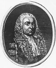
Robert Hooke, who coined the term "cell".
The first approaches to the study of cell emerged in the seventeenth century, following the development in the late sixteenth century the first microscopes. These allowed for numerous comments which have resulted in just two hundred years to a knowledge morphologically relatively acceptable. The following is a brief chronology of these discoveries:
1665: Robert Hooke published the results of his observations on plant tissues, such as cork, made with a microscope of 50x built by himself. This was the first researcher who, seeing in those tissues that were repeated units by way of cells of a honeycomb, christened as elements of repetition, "cells" (from Latin cellulae, cells). But Hooke could only watch as dead cells could not describe the structures within.
Decade 1670: Anton Van Leeuwenhoek, noted various eukaryotic cells (such as protozoa and spermatozoa) and prokaryotes (bacteria).
1745: John Needham described the presence of 'animalcules' or 'infusoria' it was single-celled organisms.
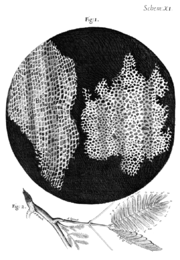 Drawing of the structure of cork observed by Robert Hooke under his microscope and as published in Micrographia
Decade 1830: Theodor Schwann cell studied animal, along with Matthias Schleiden postulated that cells are the basic units in the formation of plants and animals that are the foundation of the life process.
1831: Robert Brown described the cell nucleus.
1839: Purkinje observed the cytoplasm cell.
1850: Rudolf Virchow postulated that all cells come from other cells.
1857: Kölliker identified the mitochondria.
1860: Pasteur conducted many studies on the metabolism of yeast and the asepsis.
1880: August Weismann found that existing cells and molecules share structural similarity with cells of ancient times.
1931: Ernst Ruska built the first transmission electron microscope at the University of Berlin. Four years later, he obtained a resolving power double that of the optical microscope.
1981: Lynn Margulis published his hypothesis on the serial endosymbiosis, which explains the origin of the eukaryotic cell.
Cell theory
The concept of cell as the anatomical and functional organizations emerged between 1830 and 1880, although it was in the seventeenth century when Robert Hooke first described the existence of them, noting in a preparation plant the presence of a structure organized architecture derived from plant cell walls. In 1830 there were now microscopes with advanced optics, which allowed researchers and Theodor Schwann and Matthias
Schleiden define the principles of the cell theory, which states, inter alia:That the cell is a morphological unit of all living things: namely, that all living things are composed of cells or their secretion products.
This first assumption would be completed by Rudolf Virchow with the statement Omnis cellula ex cellula, which indicates that every cell is derived from a previous cell (biogenesis). In other words, this postulate is the refutation of the theory of spontaneous generation or de novo, which hypothesized the possibility that life was generated from inanimate elements.
A third postulate of the cell theory states that the vital functions of organisms occur within cells or in their immediate environment, and are controlled by substances they secrete. Each cell is an open system that exchanges matter and energy with their environment. Occur in a cell all vital functions, so one of them enough to have a living (which is a living being unicellular). Thus, the cell is the physiological unit of life.
Finally, the fourth postulate of the cell theory states that each cell contains all the hereditary information necessary for control of its own cycle and the development and operation of an institution of its kind, and to transmit that information to the following cell generation.
Definition
Therefore, we can define the unit cell as the morphological and functional of all living things. In fact, the cell is the smallest element that can be considered alive. As such it has a membrane of phospholipids selectively permeable maintaining an internal environment highly ordered and distinct from the external environment in terms of its composition, subject to homeostatic control, which consists of biomolecules and some metals and electrolytes. The structure is self-maintained actively through the metabolism, ensuring coordination of all cellular elements and their perpetuation through replication through a genome encoded by nucleic acids. That part of biology that deals with it is the cytology.
Features
The cells, as systems thermodynamic complexes have a set of common structural and functional elements that enable its survival, however, different cell types present modifications of these common features that allow for functional specialization and therefore gain complexity. Thus, the cells remain highly organized at the expense of increasing the entropy of the surroundings, one of the conditions of life.
Structural Features

The existence of polymers such as cellulose in plant wall cell structure allows support using an external frame.
Individuality: All cells are surrounded by an envelope (which may be a lipid bilayer naked in animal cells, a wall polysaccharide in fungi and plants, one outer membrane and other elements that define a complex wall in bacteria Gram negative; a wall of peptidoglycan, the bacterial Gram-positive, or a wall of varying composition, in archaea) that separates them and communicates with the outside, which controls cell movements and maintaining the membrane potential.
They contain an internal environment the aqueous cytosol, which forms the bulk of cell volume and they are currently in the cellular organelles.
They have shaped the genetic material DNA, the hereditary material of genes and contains the instructions for cell function and RNA, so that the former is expressed.
They have enzymes and other proteins, that support, along with other biomolecules, a metabolic active.
Functional characteristics

The enzyme, a type of proteins involved in cell metabolism.
Living cells are a complex biochemical system. The features that distinguish the cells from nonliving chemical systems are:
Nutrition. The cells take up substances from the environment, transform from one form to another, releasing energy and eliminate waste products through the metabolism.
Growth and multiplication. The cells are capable of directing its own synthesis. As a result of nutritional processes, a cell grows and divides, forming two cells in a cell identical to the original cell, through cell division.
Differentiation. Many cells can undergo changes in form or function in a process called cell differentiation. When a cell differentiates, some substances are formed or structures that were not previously trained and others that were no longer formed. Differentiation is often part of the cell cycle in which cells are specialized structures related to reproduction, dispersal or survival.
Signaling. Cells respond to chemical and physical stimuli from the external environment as much of its interior and in the case of mobile units, to certain environmental stimuli or in the opposite direction through a process called synthesis. Moreover, frequently the cells can interact or communicate with other cells, usually by means of signals or chemical messengers such as hormones, neurotransmitters, growth factors ... multicellular beings in complex processes of cellular communication and signal transduction.
Evolution. Unlike the inanimate structures, unicellular and multicellular organisms evolve. This means that there are heritable changes (which occur at low frequency in all cells on a regular basis) that may influence the overall adaptation of the cell or the upper body in a positive or negative. The result of evolution is the selection of those organisms best adapted to live in a particular environment.
The cellular properties need not be constant throughout the development of an organism: obviously, the pattern of gene expression varies in response to external stimuli, as well as endogenous factors. An important aspect to control is the pluripotency characteristic of some cells that allows them to direct their development into a range of possible cell types. In metazoans, the genetics underlying the determination of the fate of a cell is the expression of specific transcription factors specific cell lineage to which will belong, as well as epigenetic modifications. Moreover, the introduction of other transcription factors through genetic engineering in somatic cells is sufficient to induce pluripotency above, then this is one of its molecular basis.
Size, shape and function

Comparison of size between neutrophils, blood cells eukaryotes (largest), and bacteria Bacillus anthracis, prokaryotes (smaller, rod-shaped).
The size and shape of cells depends on its peripheral elements (eg wall, if any) and their internal scaffolding (ie, the cytoskeleton). Moreover, competition for space leads to a morphology characteristic tissue: for example, plant cells, polyhedral in vivo, tend to be spherical in vitro. Even there may be simple chemical parameters such as concentration gradients of salt, determining the appearance of a complex shape.
As for size, most cells are microscopic, ie are not observable to the naked eye. Despite being very small (one cubic millimeter of blood may contain about five million cells) the cell size is highly variable. The smallest cell observed under normal conditions, corresponding to Mycoplasma genitalium, 0.2 μm, being near the theoretical limit of 0.17 μm. There are bacteria with 1 and 2 μm in length. Human cells are highly variable with erythrocytes of 7 microns, hepatocytes with 20 microns, sperm of 53 μm, eggs of 150 μm and even some neurons of around one meter. In plant cells the grains of pollen may reach 200 to 300 μm and some bird eggs may reach 1 (quail) and 7 cm (ostrich) in diameter. For cell viability and function properly you should always take into account the surface-volume ratio. can substantially increase the volume of the cell and not its exchange membrane surface making it difficult level and regulation of exchange of vital substances in the cell.
Regarding its form, the cells are extremely variable, and even some do not have definite or permanent. They can be: fusiform (spindle shaped), stellate, prismatic, flattened, elliptical, globose or rounded, and so on. Some have a rigid wall and not others, allowing them to deform the membrane and cytoplasmic issue (pseudopods) to navigate or find food. There are no free cells such displacement structures but possess cilia or flagella, which are structures derived from a cellular organelle (the centrosome) that endows these cells to move. Thus, there are many cell types, related with the role, for example:
Contractile cells which are often elongated, as the muscle fibers.
Cells with thin extensions, such as neurons that transmit nerve impulses.
Cells with microvilli or folds, such as the intestine to increase the area of contact and exchange of substances.
Cuboidal, prismatic or flattened as the epithelial lining surfaces such as paving slabs.

Like it on Facebook, Tweet it or share this article on other bookmarking websites.